 English
 فارسی
فارسی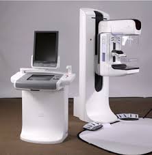
Mammography
Mammography is the process of using low-energy X-rays (usually around 30 kVp) to examine the human breast, which is used as a diagnostic and screening tool. The goal of mammography is the early detection of breast cancer, typically through detection of characteristic masses and/or microcalcifications.
Like all X-rays, mammograms use doses of ionizing radiation to create images. These images are then analyzed for any abnormal findings. It is normal to use lower-energy X-rays (typically Mo-K) than those used for radiography of bones. Ultrasound, ductography, positron emission mammography (PEM), and magnetic resonance imaging (MRI) are adjuncts to mammography. Ultrasound is typically used for further evaluation of masses found on mammography or palpable masses not seen on mammograms. Ductograms are still used in some institutions for evaluation of bloody nipple discharge when the mammogram is non-diagnostic. MRI can be useful for further evaluation of questionable findings as well as for screening pre-surgical evaluation in patients with known breast cancer to detect any additional lesions that might change the surgical approach, for instance from breast-conserving lumpectomy to mastectomy. Other procedures being investigated include tomosynthesis.
For the average woman, the U.S. Preventive Services Task Force recommended (2009) mammography every two years in women between the ages of 50 and 74.
Procedure
During the procedure, the breast is compressed using a dedicated mammography unit. Parallel-plate compression evens out the thickness of breast tissue to increase image quality by reducing the thickness of tissue that x-rays must penetrate, decreasing the amount of scattered radiation (scatter degrades image quality), reducing the required radiation dose, and holding the breast still (preventing motion blur). In screening mammography, both head-to-foot (craniocaudal, CC) view and angled side-view (mediolateral oblique, MLO) images of the breast are taken. Diagnostic mammography may include these and other views, including geometrically magnified and spot-compressed views of the particular area of concern. Deodorant, talcum powder or lotion may show up on the X-ray as calcium spots, and women are discouraged from applying these on the day of their exam. There are two types of mammogram studies: screening mammograms and diagnostic mammograms. Screening mammograms are performed yearly on a patient who presents with no symptoms and consists of only four standard X-ray images. Diagnostic mammograms are reserved for patients with breast symptoms, changes, or abnormal findings seen on their screening mammogram. Diagnostic mammograms are also performed on patients with breast implants, breast reductions, and patients with personal and/or family history of breast cancer.