 English
 فارسی
فارسی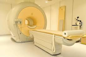
Magnetic resonance imaging (MRI) is a method of obtaining images of the interiors of objects, especially living things such as humans and animals. It does not use ionizing radiation such as X-ray s. Instead, it employs radio-frequency (RF) waves and intense magnetic field s to excite atom s in the object under evaluation. Patterns in this excitation are observed on a display. MRI can provide real-time, three-dimensional views of body organs, muscles, and joints without invasive surgery.
The MRI procedure is considered indispensable by many physicians, especially for the evaluation of sports-related injuries and for the diagnosis of chronic disease conditions. In order to properly interpret the display of an MRI, the expertise of a physician or radiologist is required. Mostly, people lack the medical knowledge to properly interpret an MRI. An MRI can reveal minor damage to tendons, ligaments, and muscles. An MRI display of the heart and surrounding arteries can provide early warning of advancing coronary disease, and can help locate cancerous tumors.
The magnetic resonance phenomenon was first demonstrated in 1946 by Felix Bloch and Edward Purcell. For the first two decades after its discovery, the phenomenon was used only for the analysis of inanimate matter. It was not until the 1990s that MRI was used to map the human brain. The science of MRI is still considered to be in its infancy. Suggested future applications include the diagnosis and treatment of as-yet unknown disease conditions and psychiatric disorders. Even behavior modification, lie detection, and thought control have been discussed as potential indirect applications of real-time MRI.
MRI is sometimes called nuclear magnetic resonance imaging (NMRI) because it involves the nuclei of atoms. However, because "nuclear" bears some negative connotations, that adjective is usually omitted.